
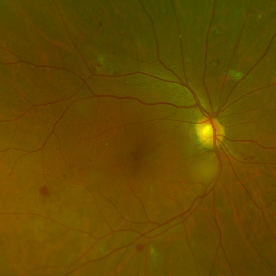
AI-assisted pre-screening · Retinal clinical trials

Enrich your retinal trial before the screening visit.

enrolled.ai grades retinal imagery against your protocol's eligibility criteria — surfacing likely-eligible candidates for reading-center confirmation, so screening visits convert instead of failing.

Built for trial sponsors, reading centers, and ophthalmology researchers.

Why pre-screen

Screen failures are the quiet budget killer in retinal trials.

Eligibility is graded, not guessed

Retinal trial protocols specify narrow ocular inclusion criteria — DRSS ranges in diabetic retinopathy studies, for example. Most referred patients fall outside them — and each one still costs a full screening visit and a reading-center grade to find out.

Manual pre-reads don't scale

Asking investigators or graders to eyeball every candidate's imagery adds weeks of queue time and pulls expert attention away from protocol reads.

Enrollment windows are short

Sites compete for the same patients. Knowing who is likely in-window before scheduling lets coordinators prioritize the right referrals first.

How it works

From image to enrichment signal in three steps.

  1. 01

    Upload retinal imagery

    Fundus photography and OCT volumes in standard formats — DICOM, TIFF, PNG, JPEG — as well as proprietary device exports. Every image is versioned and attached to the patient's FHIR record.

  2. 02

    Automated AI grading

    Our models identify and quantify relevant biomarkers and produce protocol-relevant model outputs — such as a DRSS grade for diabetic retinopathy — each with a calibrated confidence estimate. Results are written back as structured FHIR observations — traceable, queryable, exportable.

  3. 03

    Reading-center confirmation

    Flagged candidates enter the grading worklist with the AI assessment alongside the source imagery. Certified graders confirm or override — every decision captured in the audit trail.

For reading centers

A workflow that respects the grade.

enrolled.ai doesn't replace the reading center — it feeds it better. Pre-screening concentrates grader time on candidates who are plausibly in-window, while the platform keeps the full record of what the model said, what the grader decided, and when.

Reports are generated as clean, printable PDFs with the imagery, model outputs, and provenance on one page — ready for sponsor files and site records.

  • Grading worklists

    Queue of AI-flagged studies with model output, confidence, and laterality at a glance.

  • Side-by-side review

    Source imagery and model output together — confirm or override in one screen.

  • Complete audit trail

    Every classification, review, and report is an immutable, timestamped FHIR resource.

  • One-page PDF reports

    Patient, imagery, model outputs, and provenance — formatted for regulatory files.

Platform

Clinical-grade plumbing, standards first.

FHIR-native

Built on the HL7® FHIR® R4 data model. Patients, imaging studies, and AI results are standard resources your systems can already speak to.

Ophthalmic formats

DICOM, TIFF, PNG, and JPEG, plus proprietary device exports from ultra-widefield fundus and OCT systems — processed without lossy conversion.

Auditable by design

Model versions, classifications, and grader decisions are recorded as structured, timestamped events — nothing happens off the record.

Access-controlled

Project-scoped access policies separate sponsors, sites, and reading centers while sharing one source of truth.

See your screening funnel before you pay for it.

Walk through the platform with us on your own imagery, or ours.

Request a demo